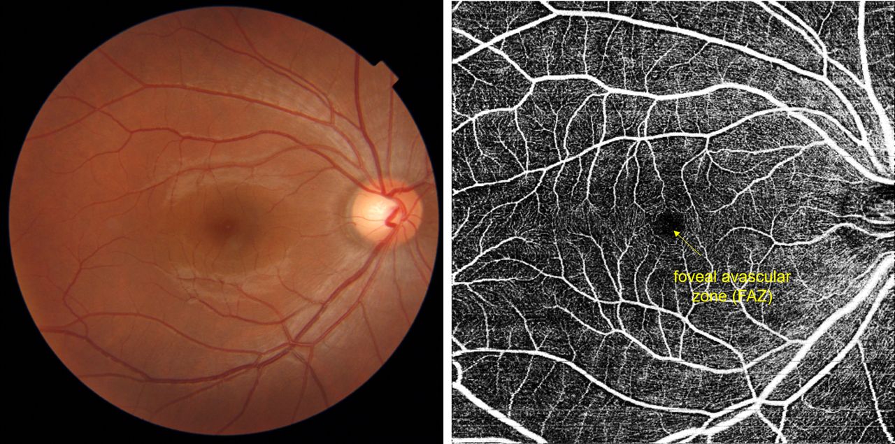
Advanced imaging of the retina provides early warning of the presence of inflammatory and neurodegenerative disorders in patients – a just-reported study finding that supports the Mind-Eye Institute’s contention the retina serves as a “poor man’s MRI.”
Scientists often refer to the retina as a “window to the brain.” That is because the retina is composed of brain tissue and tells us much about a person’s overall health, says optometrist Deborah Zelinsky, OD, in referring to the study, presented online in an April 2022 edition of Eye.
“The retina reflects what is occurring in the brain,” emphasizes Dr. Zelinsky, an internationally recognized retinal expert and the founder and research director of the Mind-Eye Institute in Northbrook, Illinois. “Eye care practitioners may someday become the professionals patients seek first for diagnosis of a variety of disorders, including early onset Alzheimer’s disease, Parkinson’s disease, high blood pressure, cardiovascular issues, and diabetes,” Dr. Zelinsky predicts.
In the Eye journal study, scientists from The Royal College of Ophthalmologists report that optical coherence tomography (OCT), an advanced technology providing detailed resolution of the retina and optic nerve, easily “visualizes” structural and neurological changes – retinal “biomarkers” — associated with inflammatory and neurodegenerative diseases. OCT could prove to be a “non-invasive way to monitor the status of the [patient’s] central nervous system” and facilitate multidisciplinary approaches to treating such disorders, they state.
“The retina and optic nerve are considered extensions of the central nervous system (CNS) and thus can serve as the window for evaluation of CNS disorders,” the researchers write, echoing Dr. Zelinsky’s own sentiments as presented in her publications and scientific addresses.
Dr. Zelinsky also references a study published earlier this year (2022) in Nature Reviews Neurology. In it, researchers conclude that OCT is useful for the early diagnosis and management of Parkinson’s disease, a neurodegenerative brain disorder. “Visual disturbances observed in patients with Parkinson’s disease are linked to retinal dopamine loss,” study authors write. Dopamine is a neurotransmitter. A deficiency in dopamine can interfere with coupling and interaction among retinal cells, Dr. Zelinsky says.
The Mind-Eye Institute has long been applying the principles of advanced optometric science to stimulate the retina in order to modify the dynamic relationship between the mind’s visual inputs and the body’s internal responses. Retinal stimulation is achieved through use of highly individualized, therapeutic eyeglasses, filters, and other optometric tools that can vary the amount, intensity and angle of light passing through the retina into the optic nerve.
Environmental signals in the form of light enter the retina and are converted into electrical signals. Those signals propagate through neurons and interact with critical brain structures. Retinal signals affect not just the visual cortex for eyesight, but other, significant regions of the brain as well. Retinal stimulation using variances in light can promote positive (or negative) alterations in a person’s basic physical, physiological, and even psychological systems involved in circuitry of motor control, posture, emotion, and thinking, Dr. Zelinsky explains.
“The latest studies clearly indicate retinal changes mirror degenerative changes in the brain and suggest retinal neuromodulation of brain activity through non-invasive stimulation of the eye — alone or in combination with other therapies – could prove clinically effective in addressing a host of neurodegenerative disorders and mental illnesses like schizophrenia,” Dr. Zelinsky states.

Research, ADD/ADHD/Learning Disorders, Auditory Processing, Autism Spectrum Disorder, TBI/Concussion/Stroke, Visual Processing
Not Surprisingly, Retina Offers Early Warning of Disease
Latest Study Underscores Mind-Eye Founder’s Longtime Contention
