Could changes in the retina be an early warning sign of developing Alzheimer’s disease? Recent studies indicate yes, including research just published in a May 2023 issue of the journal Alzheimer’s and Dementia. Even more exciting, the latest findings suggest that, some day in the relatively near future, the retina might be used or stimulated as a way of mitigating symptoms of Alzheimer’s and other neurodegenerative disorders.
Why may all this be possible? Because the retina, which is composed of brain tissue and plays a critical role as part of the central nervous system, is the only part of the brain that is exposed and readily accessible to non-invasive techniques, such as stimulation with light.
As I have frequently pointed out, the Mind-Eye Institute is internationally known for its use of therapeutic eyeglasses and other advanced optometric tools to manipulate the amount, angle, and intensity of light passing through the retina. Those changes in lighting can promote the development of new brain pathways in patients struggling with the symptoms from traumatic head injury, concussion, stroke, and neurological disorders. Retinal stimulation has also proven effective in building undeveloped visual processing skills in children – and adults – with autism spectrum disorder, attention deficit hyperactivity disorder, and other learning, socialization, and behavioral difficulties.
Environmental signals (in the form of light) enter the retina and convert into electrical signals, which propagate through neurons and interact with key brain structures. These retinal signals affect not only the visual cortex but other, significant regions of the brain as well, linking with structures like the limbic system, the cerebellum, and brainstem.
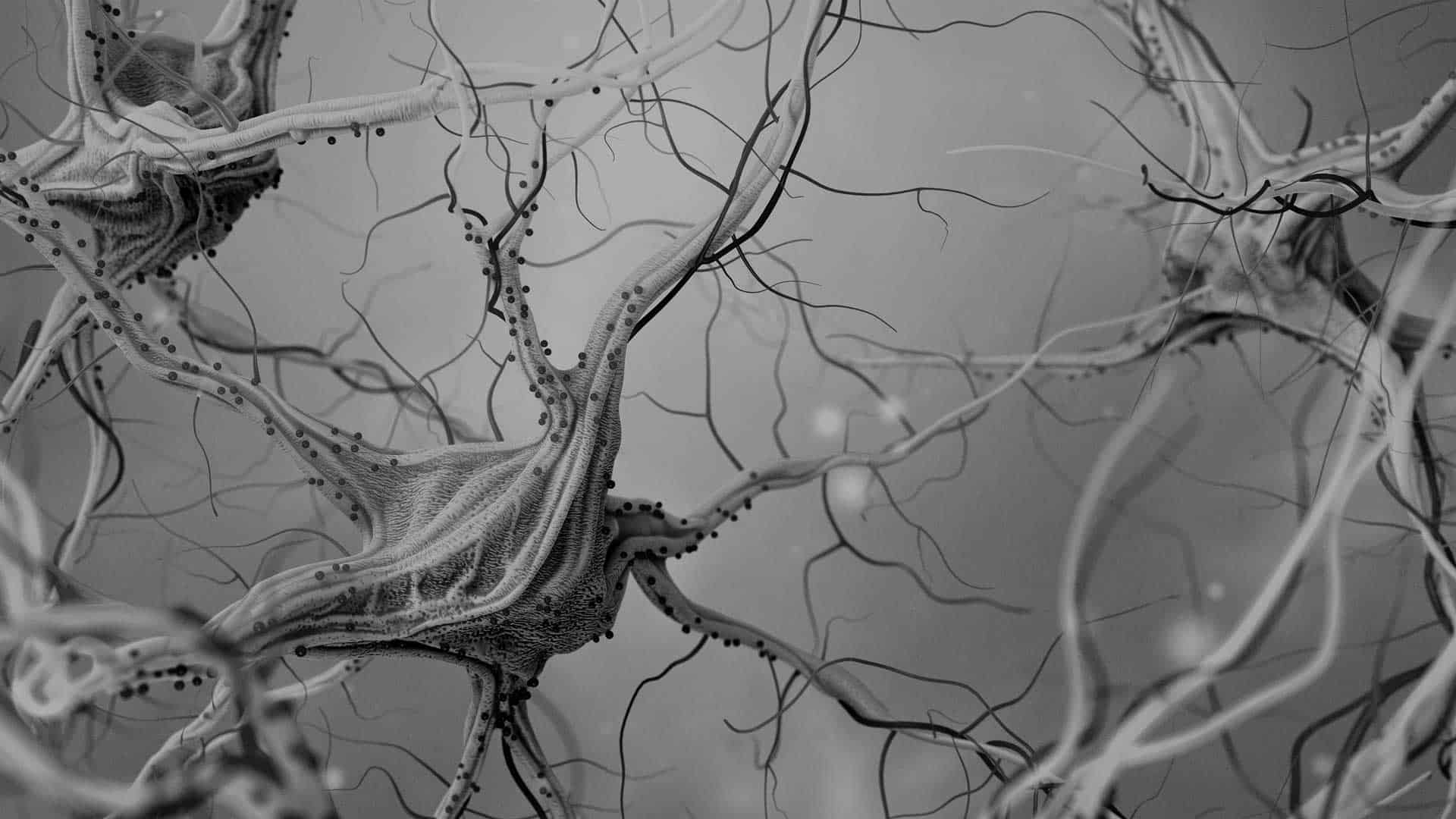
The retina is a window to the brain. The latest science confirms this fact and says retinal degeneration reflects and parallels similar degeneration occurring in the brain. Retinal changes mirror brain changes in patients with Alzheimer’s disease, multiple sclerosis, schizophrenia, and Parkinson’s. That is why, as scientists and researchers, we believe neuromodulation of brain activity through non-invasive, light stimulation of the eye -- alone or in combination with other therapies -- will eventually prove clinically effective in addressing a host of neurodegenerative diseases, mental illnesses, autism, metabolic disorders, and dysfunctions in circadian rhythm and endocrine systems.
What is interesting about the most recent study in Alzheimer’s and Dementia is the finding that detectable abnormalities in the retina’s blood vessels – specifically, vascular disruptions that restrict blood flow and allow toxic substances to pass the blood-retinal barrier and contaminate the retina -- are potential harbingers of developing Alzheimer’s disease. Accumulations of the protein beta-amyloid can clog small blood vessels and arteries in the retina. This waste protein is also associated with Alzheimer’s. Study investigators determined that, in patients with Alzheimer’s disease or other types of mild cognitive impairment, as much as 70 percent of the blood-retinal barrier may be damaged.
Alzheimer’s disease is a progressive, neurodegenerative disorder that leads to cognition impairment and irreversible memory loss. It is the most common form of dementia and afflicts about 10 percent of the population over age 65.
If not flushed from the brain through the glymphatic system, beta-amyloids begin to clump together, forming plaque that impairs communication among brain cells and leads to progression of Alzheimer’s disease. The glymphatic system is a pathway, activated during sleep, which allows cerebral spinal fluid to enter the brain and clear it of waste products, including beta-amyloid.
Until recently, amyloid buildup could only be determined by analyzing the brains of Alzheimer’s patients post-mortem. However, recent investigations show amyloid accumulation is measurable outside the brain -- in retinal and optic nerve locations -- and linked to malfunctioning circadian and cognitive circuitry in the brain. Circadian rhythm is disrupted when specific retinal cells involved with sleep become target of amyloid plaques. Also, cognitive changes are further evidenced by thinning of retinal tissue that corresponds to the brain’s hippocampal volume.
Another recent study, in a 2022 issue of the Journal of Cell Immunology, reports that “diminished uptake of glucose in the brain is a better marker for classifying Alzheimer’s disease than beta-amyloid…deposition.” Other research has shown that this glucose metabolism can be changed or regulated by using light to stimulate the retina.
So, what is the significance of these findings for the millions of patients with Alzheimer’s disease or other forms of dementia?
As the Alzheimer’s Association emphasizes, early detection and diagnosis are critical factors in providing patients with proper treatment to help mitigate symptoms and, perhaps, slow progression of disease. Ongoing advancements in retinal imaging eventually will allow optometrists and other eye specialists to detect brain changes at an earlier point when symptoms are only mild or, perhaps, not yet even manifest and, therefore, more responsive to therapy.
As I have said in the past, optometric science is moving well beyond what one might consider its horse-and-buggy days of the 19th and 20th centuries when the main goal was simply to give patients central eyesight of 20/20 acuity. Advancements in our understanding of the retina, and the technology required to evaluate it could one day make optometrists the go-to professionals for early detection and diagnosis of a host of serious disorders.
The longtime motto of the Mind-Eye Institute is: Let’s leave 20/20 in the 20th Century. Why not add to that: “Let’s build better brains in the 21st Century!”
Deborah Zelinsky, O.D.
Founder, Executive Research Director
The Mind-Eye Institute
Northbrook, Illinois

